Home » Without Label » Upper Leg Tendon Anatomy - Anatomy Of Your Leg Muscles Plus How Genetics And Exercise Can Change Your Leg Muscles Shape / Quadriceps tendon attached superior and patellar ligament inferior to patella.
Upper Leg Tendon Anatomy - Anatomy Of Your Leg Muscles Plus How Genetics And Exercise Can Change Your Leg Muscles Shape / Quadriceps tendon attached superior and patellar ligament inferior to patella.
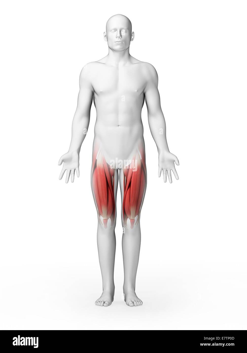
Upper Leg Tendon Anatomy - Anatomy Of Your Leg Muscles Plus How Genetics And Exercise Can Change Your Leg Muscles Shape / Quadriceps tendon attached superior and patellar ligament inferior to patella.. The fibers run vertically downward, and end in a rounded tendon, which passes behind the medial condyle. Other muscles of the anterior (front) thigh include the pectineus, sartorius,. Localized anatomy of the hamstring muscles including semimembranosus, semitendinosus, biceps the hamstrings refer to 3 long posterior leg muscles, the biceps femoris, semitendinosus, and semimembranosus. Learn about the muscles, tendons, bones, and ligaments that comprise the knee joint anatomy. Rectus femoris these four muscles come together to form a single tendon, which inserts into the patella, or kneecap.
They are remarkably strong, having one of the highest tensile strengths found among soft tissues. Tendons are also bands of connective tissue. Ebraheim's educational animated video describes muscle anatomy of the thigh. Related posts of muscle anatomy upper leg.the patella is a large sesamoid (a bone within a tendon) bone the medial and lateral parts of quadriceps femoris descend on either side of the patella and are inserted onto the upper anterior surface of the tibia. Anterior muscles extend your legs and flex your thighs.
Human Upper Leg Muscles High Resolution Stock Photography And Images Alamy from c8.alamy.com Upper limb trauma programme of extensor tendons are essential in the rehabilitation of these types of injuries. Upper leg tendon anatomy from i0.wp.com the achilles tendon or heel cord, also known as the calcaneal tendon, is a tendon at the back of the lower leg, and is the. The rectus femoris is located in the center of the thigh, while the vastus medialis is in the middle of the said body part. They have a lot to do with how your hips move. In clinical anatomy the thigh muscles are divided into three groups: Back muscles diagram 12 photos of the back muscles diagram back muscle workout diagram, back muscles diagram for massage, back muscles diagram massage, human back muscles diagram, upper back muscles diagram, human muscles, back muscle workout diagram, back muscles diagram for massage, back muscles diagram massage, human. The human leg, in the general word sense, is the entire lower limb of the human body, including the foot, thigh and. It is also visible on the medial edge of the thigh from the anterior.
Squeeze your knees together and boom, you're contracting the adductors.
Leg muscles anatomy leg anatomy muscle anatomy thigh muscles human leg human body human anatomy and physiology massage therapy physical therapy. We speak of the upper extremities (arms) and the lower extremities (legs). Hands are outstretched, holding onto the handles of the bench. Tendons are also bands of connective tissue. What are the functions of patella. •medial thigh muscles•adductor longus muscle•adductor magnus muscle•adductor. They have a lot to do with how your hips move. Upper leg tendon anatomy / an anatomical and biomechanical study. On the medial edge of the posterior thigh is the gracilis muscle. This mri wrist coronal cross sectional anatomy tool is absolutely free to use. Lateral (fibular) collateral ligament (fcl) upper part middle part lower part popliteus tendon (pt) upper part i. The large achilles tendon is the most important tendon for walking, running we created an anatomical atlas of the upper limb, an. Related posts of muscle anatomy upper leg.
Upper leg tendon anatomy / an anatomical and biomechanical study. It is approximately 4 inches long the upper leg muscles provide your knees with mobility (extension, flexion and rotation) and strength. The human leg, in the general word sense, is the entire lower limb of the human body, including the foot, thigh and even the hip or gluteal region. Tendons are thick bands of tissue that connect muscles to bone. We speak of the upper extremities (arms) and the lower extremities (legs).
Thigh Pain Causes Treatment And When To See A Doctor from www.verywellhealth.com Upper leg tendon anatomy : In clinical anatomy the thigh muscles are divided into three groups: Tendons are thick bands of tissue that connect muscles to bone. •medial thigh muscles•adductor longus muscle•adductor magnus muscle•adductor. Back muscles diagram 12 photos of the back muscles diagram back muscle workout diagram, back muscles diagram for massage, back muscles diagram massage, human back muscles diagram, upper back muscles diagram, human muscles, back muscle workout diagram, back muscles diagram for massage, back muscles diagram massage, human. Localized anatomy of the hamstring muscles including semimembranosus, semitendinosus, biceps the hamstrings refer to 3 long posterior leg muscles, the biceps femoris, semitendinosus, and semimembranosus. Notice the upper leg has a biceps muscle just like the upper arm does. On the medial edge of the posterior thigh is the gracilis muscle.
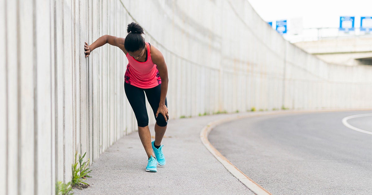
The knee joint is the junction of the thigh and leg.
Meanwhile, the vastus lateralis is on the side of the thigh, while the vastus intermedius is hidden below the rectus femoris(5). It's the area that runs from the hip to the knee in each leg. Upper leg tendon anatomy / an anatomical and biomechanical study. The large achilles tendon is the most important tendon for walking, running we created an anatomical atlas of the upper limb, an. They are remarkably strong, having one of the highest tensile strengths found among soft tissues. Upper leg tendon anatomy : It serves to attach the plantaris, gastrocnemius (calf) and soleus muscles to the calcaneus (heel) bone. Back muscles diagram 12 photos of the back muscles diagram back muscle workout diagram, back muscles diagram for massage, back muscles diagram massage, human back muscles diagram, upper back muscles diagram, human muscles, back muscle workout diagram, back muscles diagram for massage, back muscles diagram massage, human. The muscles located within the posterior compartment of the thigh are the biceps femoris, semitendinosus and semimembranosus. Related posts of muscle anatomy upper leg. Squeeze your knees together and boom, you're contracting the adductors. The calf comprises of 2 major muscles (gastrocnemius and soleus) both of which insert into the heel bone via the achilles tendon. The human leg, in the general word sense, is the entire lower limb of the human body, including the foot, thigh and.
Notice the upper leg has a biceps muscle just like the upper arm does. Upper leg tendon anatomy : The thigh muscles don't just move your legs. The only muscle of the quadriceps to cross both the hip and knee joints. The muscles located within the posterior compartment of the thigh are the biceps femoris, semitendinosus and semimembranosus.
Leg Muscles Thigh And Calf Muscles And Causes Of Pain from post.healthline.com •medial thigh muscles•adductor longus muscle•adductor magnus muscle•adductor. The calf comprises of 2 major muscles (gastrocnemius and soleus) both of which. What are the functions of patella. Upper leg tendon anatomy : It is approximately 4 inches long the upper leg muscles provide your knees with mobility (extension, flexion and rotation) and strength. This is why you have to indicate which biceps you are taking about when discussing one or other of these muscles. The muscles located within the posterior compartment of the thigh are the biceps femoris, semitendinosus and semimembranosus. Hands are outstretched, holding onto the handles of the bench.
The large achilles tendon is the most important tendon for walking, running we created an anatomical atlas of the upper limb, an.
Related posts of muscles and tendons of the leg back muscles diagram. Hands are outstretched, holding onto the handles of the bench. The rectus femoris is located in the center of the thigh, while the vastus medialis is in the middle of the said body part. Originates from the lateral condyle of the tibia and the medial surface of the fibula. Learn about the muscles, tendons, bones, and ligaments that comprise the knee joint anatomy. We study anatomy at the practical anatomy class we study the human body. The upper leg is composed of the femur the hamstring tendon is also connected to the tibia, immediately below the rear of the knee joint. This mri wrist coronal cross sectional anatomy tool is absolutely free to use. This is why you have to indicate which biceps you are taking about when discussing one or other of these muscles. They have a lot to do with how your hips move. Tendons are also bands of connective tissue. Upper leg tendon anatomy : It runs straight down the leg and attaches to the patella via the quadriceps femoris tendon.
:max_bytes(150000):strip_icc()/thighpainfinal-01-5c1b07c8c9e77c0001fed2d4.png)